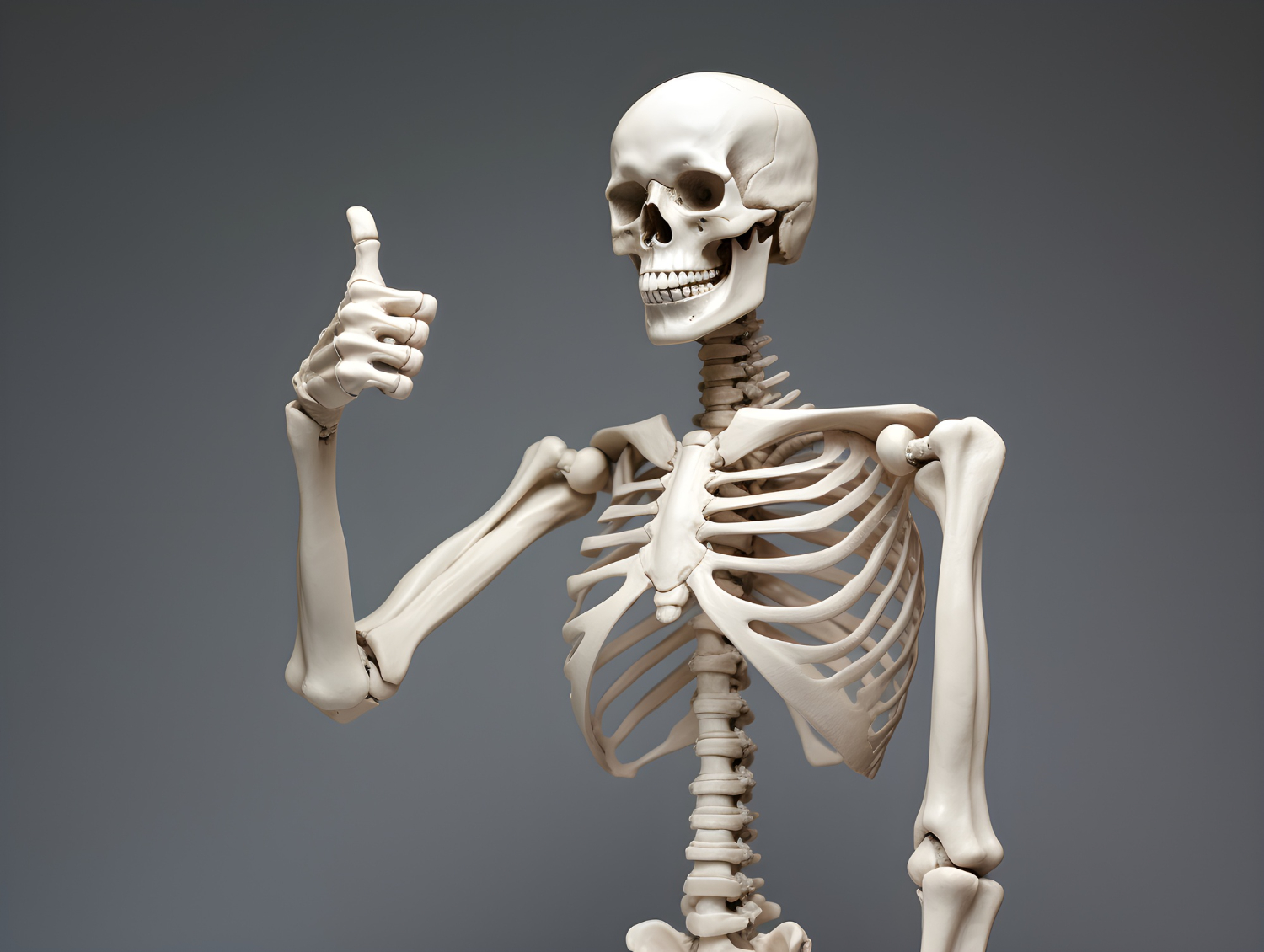
Overview of skeletal system of human. The human skeletal system is a complex framework that provides structural support, protection for internal organs, leverage for movement, storage of minerals, and production of blood cells.
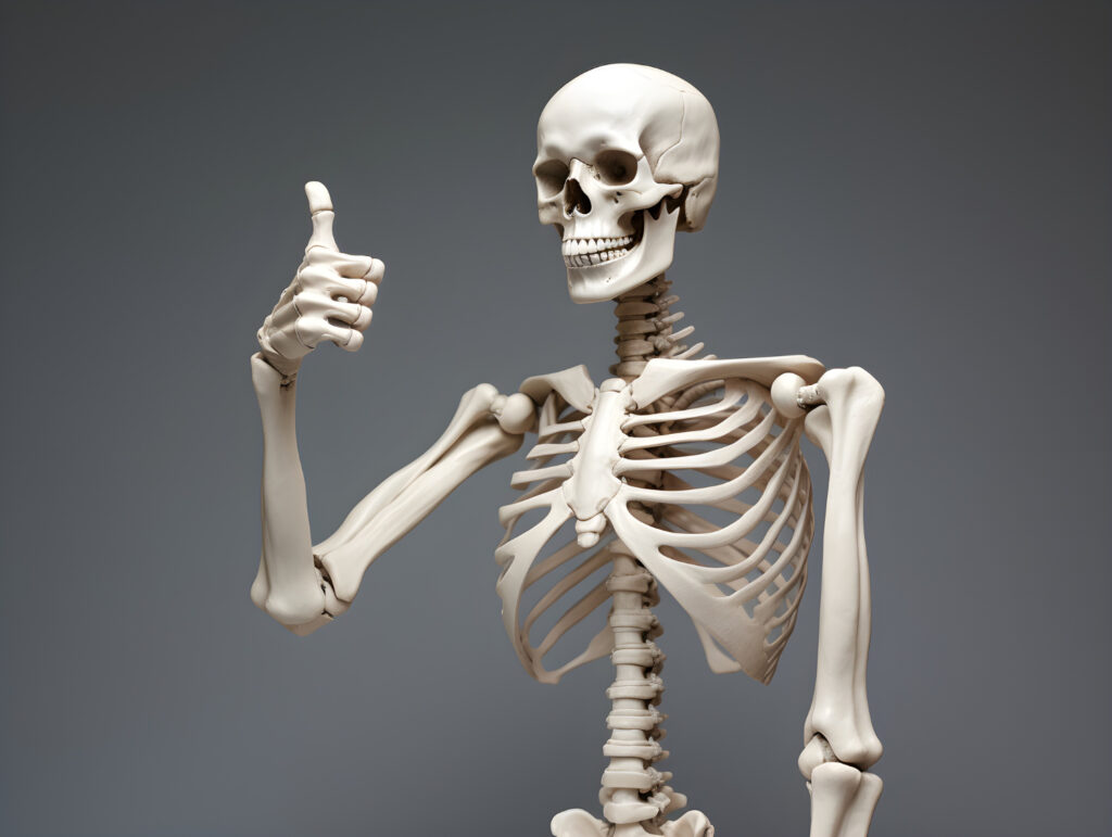
Skeletal System of Human:
(i) Axial Skeleton and (ii) Appendicular Skeleton
Axial Skeleton:
- Axial skeleton protects the internal organ lies on th central axis of the body.
- 80 bones are in axial skeleton of a human body.
- Axial Skeleton: Skull, Thoracic cage, Vertebral column
Skull:
- Skull consists of cranial bones and the facial skeleton.
- Cranial bones compose the top and back of the skull and enclose the brain.
- Facial skeleton, as its name suggests, makes up the face of the skull.
Cranial Bones :
- 8 cranial bones support and protect the brain.
- (i) Frontal bone. (ii) Parietal bone (iii) Occipital bone (iv) Temporal bone (v)Sphenoid bone (vi) Ethmoid bone
Facial bones:
- 14 bones of the facial skeleton form the entrances to the respiratory and digestive tracts.
- (i)Nasal Bone (ii) Palatine Bone (iii) Zygomatic bone (iv) Lacrimal bone (v)Inferior nasal conche (vi) Maxillae (vii) Mandible (viii) Vomero
Bone’s of the Inner Ear / Ear ossicles :
- Inside the Petrous part of the temporal bone are the three smallest bones of the body.
- (i) Mallens (ii) Incus (iii) Stapes
- These three bones articulate with each other and transfer vibrations from the tympanic membrane to the inner ear.
Laryngeal skeleton/Hyoid bone:
- Located in the anterior midline of the neck, between the chin and the thyroid cartilage.
- Its primary functions include supporting the tongue and involved in swallowing and speech production.
Skull Sutures:
In foetuses and newborn infants, cranial bones are connected by flexible fibrous sutures, including large regions of fibrous membranes called fontanelles.
- These regions allow the skull to enlarge to accommodate the growing brain.
- The sphenoidal, mastoid and posterior fontanelles close after two months, while the anterior fontanelle may exist for up to two years.
- As fontanelles close, sutures develop. Skull sutures are immobile joints where cranial bones are connected with dense fibrous tissue.
Four major cranial sutures –
- Lambdoid suture (Between Occipital and Parietal bones)
- Coronal suture (Between Frontal & Parietal bones)
- Sagittal suture (Between two Parietal bones)
- Squamous suture (Between Parietal and temporal bones)
Thoracic Cage:
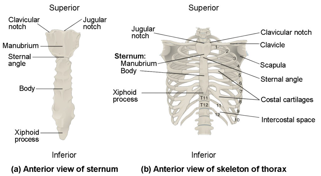
- The thoracic cage, formed by the ribs and sternum, protects internal organs and gives attachment to muscles involved in respiration and upper limb movement.
- Sternum consists of the manubrium, body of sternum and xiphoid process.
- True Ribs: Ribs 1-7 pairs are called true ribs because they they directly attached to the sternum.
- False Ribs: Ribs from 8-10 pairs are known as false ribs.
- Floating Ribs: Ribs from 11-12 pairs are called floating Ribs as they are not connected with the sternum.
Vertebral Column:
- The vertebral column is a flexible column formed by a series of 24 vertebrae, plus the Sacrum and Coccyx.
- Commonly referred as spine, the vertebral column extends from the base of the skull to the pelvis.
- The spinal cord passes from the foramen magnum of the skull through the vertebral canal within the vertebral column.
Vertebral column is grouped into five regions –
- (i) Cervical spine (C01-C07)
- (ii) Thoracic Spine (T01-T12)
- (iii) Lumbar spine (L01-L05)
- (iv) Sacral spine
- (v) Coccygeal spine.
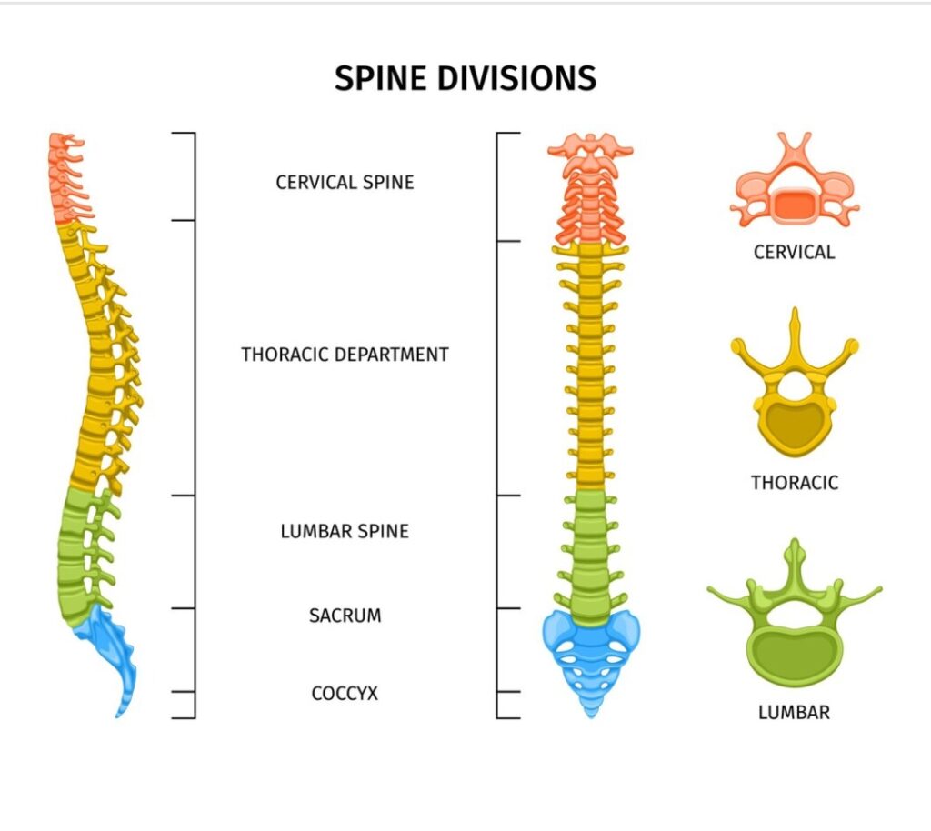
- Spine is also known as the spinal or vertebral column or simply “the backbone”. This strong yet flexible central support holds the head and torso upright yet allows the neck and back to bend and twist.
- With the S shape, it acts like a spring and can flex when we are young and jump of something.
- If it was straight up and down, it could break easily.
- Spinal joints do not have a wide range of movement but they still allow flexibility.
Appendicular Skeleton
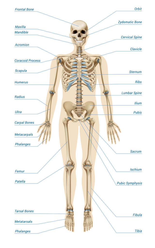
- Bones of the appendicular skeleton facilitate movements – Girdles and Limbs.
- Out of the 206 bones, 64 in the upper appendicular and 62 in the lower appendicular skeleton.
Bones of the Shoulder girdle:
- The pectoral or shoulder girdle consists of Scapula and Clavicles.
- The shoulder girdle connects the bone of the of the upper limbs to the axial skeleton.
- These bones also provide attachment for muscles that move the shoulders and upper limbs.
Bones of the upper limb:
- The upper limbs include the bones of the arm, forearm, wrist and hand.
- Humerus, Radius, Ulna, Carpals, Metacarpals & Phalanges.
- The only bone of the arm is the humerus which articulates with the forearm bones – the radius and ulna at the elbow joint.
- The ulna is the larger of the two forearm bones.
- The wrist or carpus, consists of eight carpal bones.
- Eight carpal bones of the wrist:- Scaphoid, Lunate, Triquetral, Pisiform, Trapezoid, Trapezium, Capitate and Hamate.
- 5 bones that form the palm. (metacarpals).
- 14 bones (Phalanges) that form the fingers & thumb.
Bones of the Pelvic girdle:
- The pelvic girdle is aring of bones attached to the vertebral column, that connects the bones of lower limbs to the axial skeleton.
- Pelvic girdle consists of right and left hip bones.
- Each hip bone is a large, flattened and irregulary shaped, fusion of three bones – Ilium, Ischium & Pubis
- The female pelvic brim is larger and wider than the male’s due to child bearing adaptions.
Bones of the Lower limb:
- The lower limbs Include the bones of the thigh, leg and foot.
- The femur is the only bone of the thigh. It is the largest bone in the body.
- Femur articulates with the two bones of the leg – the larger tibia and smaller fibula.
- The thigh and leg bones articulate at the knee joints that is protected and enhanced by the patella bone that supports the quadriceps tendon.
- Tarsal bones of the ankle.
- Ankle bones or tarsus consists of 7 tarsal bones: Calcaneus, Talus, Cuboid, Navicular & 3 cuneiforms.
- The arches of the foot are formed by the interlocking bones and ligaments of the foot.
- Metatarsals that form the foot’s arch.
- Phalanges that form the toes.
- They serve as shock-absorbing structures that support body weight and distribute stress evenly during walking.
- With each step, the main weight of the body moves from the rear to the front of the foot.
- The heel region bears the initial pressure as the foot is put down.
- The force passes along the arch which flattens slightly then recoils to transfer the energy and pressure to the ball of the foot, and finally to the big toe for the push-off.
Types of Bones
Bones of the human skeletal system. are categorised by their shape and function into five types.
1) Long Bone:
- Outside of long bone consists of a layer of compact bone surrounding the spongy bone.
- Example: Femur, Humerus etc.
2) Short Bone:
- Located in the wrist and ankle joints,
- Short bones provide stability and some movements.
- Example: Carpals, Tarsals etc.
3) Flat Bones:
- Function of the flat bones is to protect internal organs.
- Provide protection, like a shield and also can provide large areas of attachment of muscles.
- Example: Ribs, Sternum, Pelvic Girdle, Frontal, Parietal, Occipital, Lacrymal, Nasal, vomero bones
4) Irregular Bones:
- Irregular bones vary in shape ond structures.
- Have complex shape, which helps protects internal organs.
- Example: Vertebrae.
5) Sesamoid Bones:
- These small round bones are commonly found in tendons of the hands, knees & feet, protect from stress and wearing.
- Example: Patella (knee cap)

Functions of the Skeletal System:
- Support: Provides a rigid framework that supports the body and maintains its shape.
- Protection: Encases vital organs (e.g., skull protects the brain, rib cage protects the heart and lungs).
- Movement: Provides attachment points for muscles, allowing leverage and motion.
- Mineral Storage: Stores essential minerals, particularly calcium and phosphorus, and releases them into the bloodstream as needed.
- Blood Cell Production: Houses red marrow in certain bones, which produces red and white blood cells and platelets (hematopoiesis).
Bone Health and Disorders:
- Osteoporosis: A condition characterized by weakened bones due to loss of bone mass, increasing the risk of fractures.
- Arthritis: Inflammation of the joints causing pain and stiffness, including osteoarthritis and rheumatoid arthritis.
- Fractures: Breaks or cracks in bones resulting from trauma or stress, requiring proper medical intervention for healing.
