Answers of long question from PYQ Paper Comparative Anatomy of Semester IV of the years 2019, 2021 & 2022 respectively.
Table of Contents
Comparative Anatomy 2019
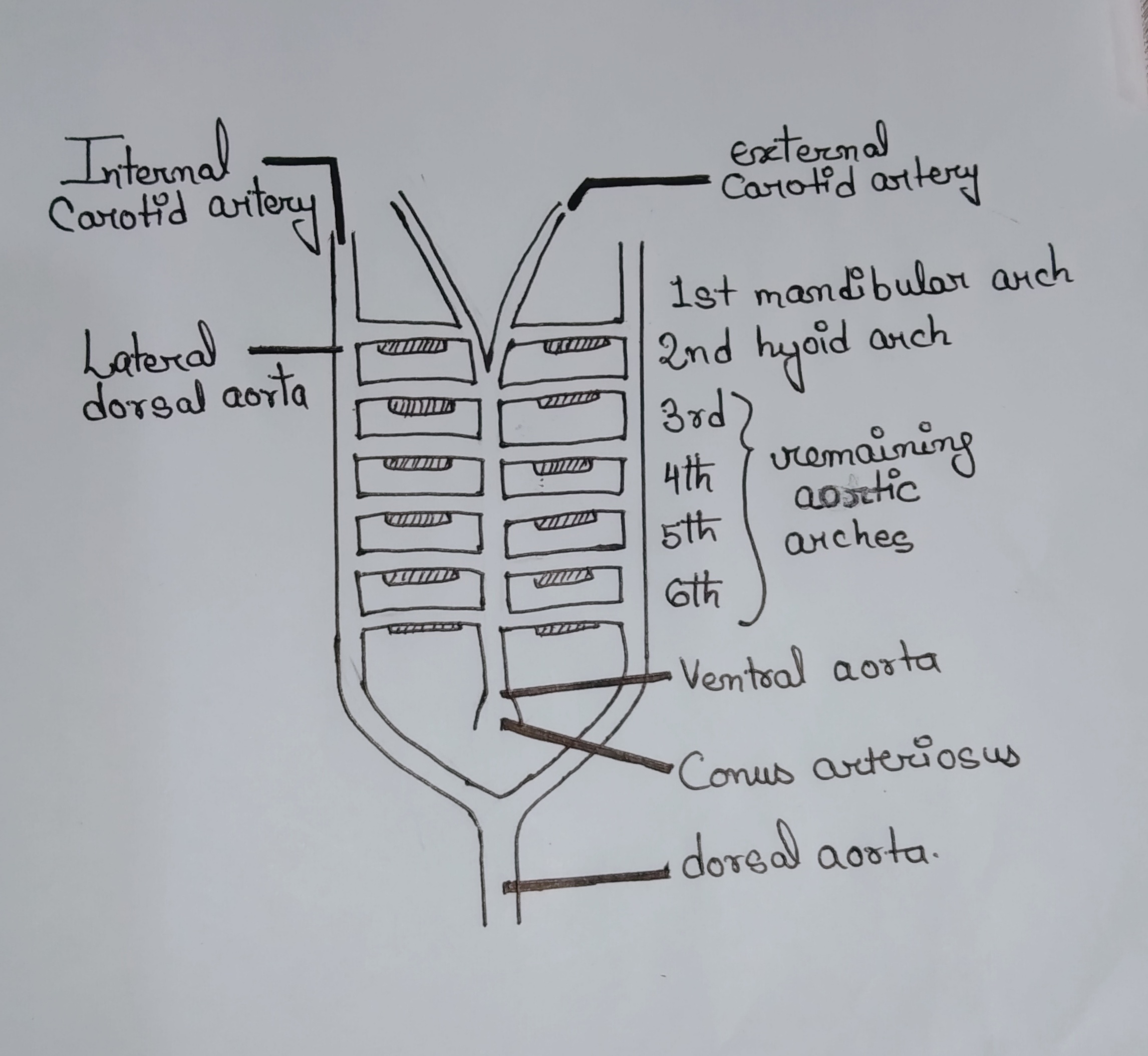
3.a) Write about a fundamental plan of the aortic arch in the vertebrate with a diagram.
Ans→ The aortic arches are the blood vessels that supply the pharyngeal arches and they serve as a communication between the ventral and dorsal aortae. They are paired, serving the left and right pharyngeal regions which are basically similar in number and disposition of different vertebrates during the embryonic stages.
The number of aortic arches is different in adult vertebrates but they are built on the same fundamental plan in embryonic life. There are 6 pairs of aortic arches developed in similar fashion from anterior to posterior region/part. The aortic arches are : Mandibular arch (I) Hyoid arch (II) III, IV, V, VI are the branchial aortic arches.
These 6 pairs of aortic arches are attached with the ventral and dorsal aortae.
There is a progressive reduction of aortic arches in the vertebrates series during evolution. The difference in the number of aortic arches are due to the complexity of heart circulation in the mode of living from aquatic to terrestrial respiration.
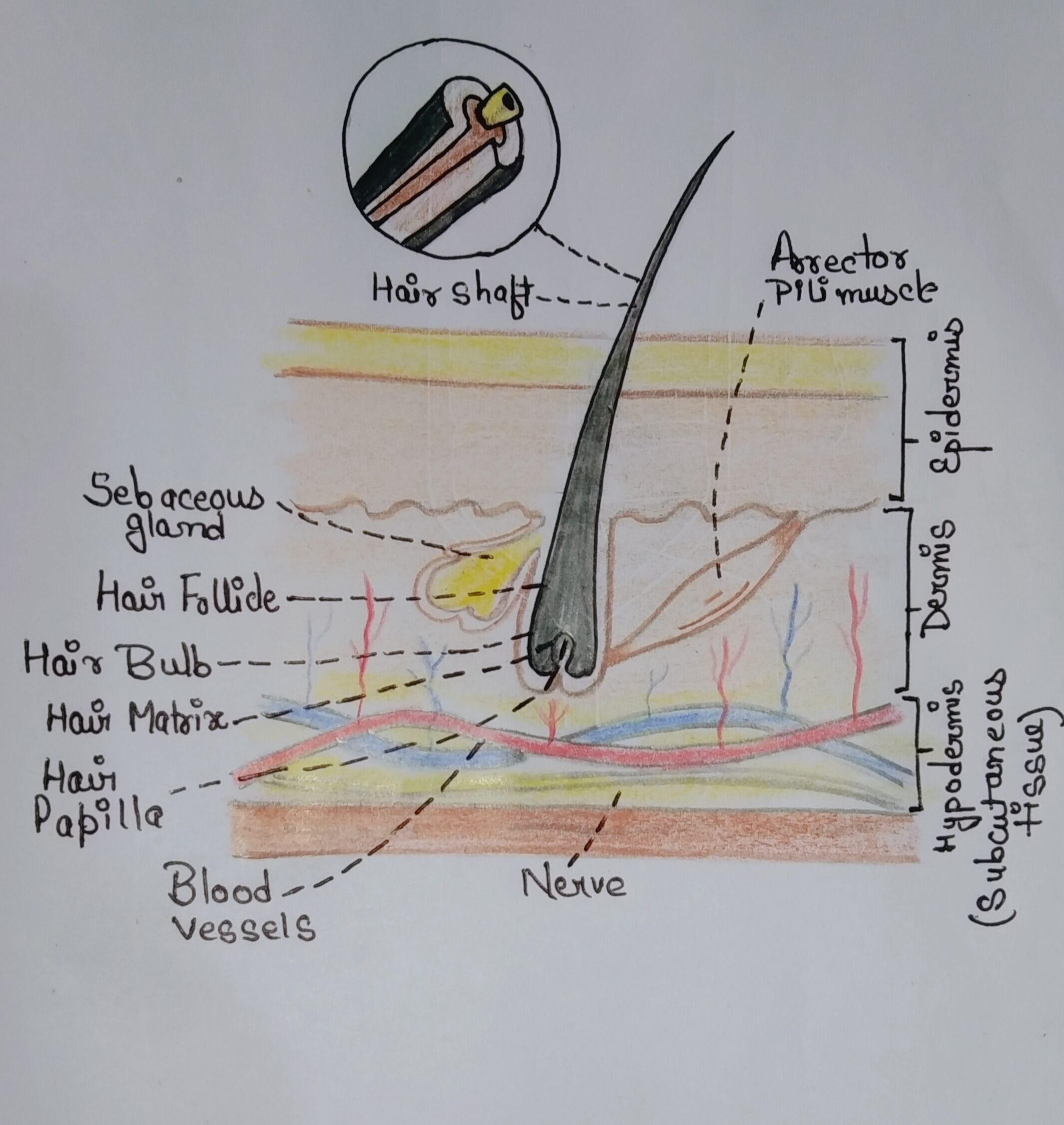
3.b) Draw and describe the microscopic structure of mammalian hair with associated glands?
Ans→Hair is a derivative of the epidermis and consists of two distinct parts- (I) Hair Shaft and (II) Hair Root
(I) Hair Shaft:
- Cuticle: Outermost layer of the hair composed of overlapping, flat, keratinized cells.
- Cortex: Middle layer of the hair contains densely packed keratin and pigment granules, giving hair its strength and colour.
- Medulla: Innermost core of the hair may be absent in fine hair, composed of loosely packed cells and air spaces.
(II) Hair Root
- Hair Follicle: The living part of the hair located under the skin.Tubular invagination of the epidermis surrounding the hair root.
- Hair Bulb: Enlarged base of the hair root and contains the dermal papilla and the matrix where hair growth begins.
- Dermal Papilla: Cone-shaped elevation at the base of the hair bulb which contains blood vessels that nourish the hair.
- Matrix: Mitotically active cells surrounding the dermal papilla which are responsible for producing new hair cells.
(III) Associated Structures
- Arrector Pili Muscle: Small muscle attached to the hair follicle; contracts to make the hair stand up (goosebumps).
1.) Sebaceous Glands: Produce sebum, which lubricates and waterproofs the hair and skin.
2.) Sweat Glands:
- Eccrine Glands: Simple coiled tubular glands that open directly onto the skin surface, distributed throughout the skin. Regulate body temperature through sweat production.
- Apocrine Glands: Larger than eccrine glands and open into hair follicles, secrete a thicker, milky fluid that can lead to body odour when broken down by bacteria.
3.c) Write down the nature, distribution and functions of the V and VIII cranial nerves in mammals. What is nervous terminalis?
Ans→ Trigeminal Cranial Nerve (V)
1.) Nature: Mixed (both sensory and motor)
2.) Distribution: Trigeminal nerve divided into three branches:
- Ophthalmic branch (V1): Sensory innervation to the forehead, scalp, and upper eyelid.
- Maxillary branch (V2): Sensory innervation to the lower eyelid, upper lip, and cheek.
- Mandibular branch (V3): Sensory and motor innervation to the lower jaw, teeth, gums, and muscles involved in chewing.
3.) Functions:
- Sensory: Sensation of touch, pain, and temperature from the face, scalp, and mouth.
- Motor: Movement of the muscles involved in mastication (chewing).
Vestibulocochlear Cranial Nerve (VIII)
1.) Nature: Sensory
2.) Distribution: Vestibulocochlear nerve divided into two branches:
- Cochlear nerve: Innervates the cochlea of the inner ear.
- Vestibular nerve: Innervates the semicircular canals, utricle, and saccule of the inner ear.
3.) Functions:
- Cochlear nerve: Helps in hearing. Transmits sound information from the cochlea to the brain.
- Vestibular nerve: Helps in balance. Transmits information about head position and movement from the vestibular apparatus to the brain.
Comparative Anatomy 2021
3.a) What do you understand by your suspension? What is autostylic jaw suspension? How an autostylic jaw suspension differs from craniostylic type of jaw suspension. Give one example of where you can find a holostylic type of jaw suspension.
Ans→ Jaw suspension is the attachment of lower jaw with the upper jaw or with skull for the efficient biting and chewing.
Autostylic Jaw Suspension: The condition to which pterygo quadrate is modified to form epipterygoid and quadrate and intimately bound to cranium by investing dermal bones (auto- self). Hyomandibular become modified into columella or stapes of middle ear for transmitting sound waves not available for jaw suspension.
Examples: Chiameres, Lungfish, Tetrapods etc.
In autostylic jaw suspension, the upper jaw is fused to the cranium, with the lower jaw attached directly to the skull. Where in craniostylic jaw suspension, the upper jaw is part of the cranium, and the lower jaw (mandible) is a separate bone, attached by a joint.
An example of holostylic jaw suspension is found in chimaeras (also known as ratfish or ghost shark
3.b) Describe ruminant stomach with a diagram.
Ans→ The ruminants swallow herbage without completing mastication. This habit enables timid herbivores to produce and swallow food at intervals that can be digested later in safer circumstances. In a typical ruminant such as sheep, cow etc. the stomach is complicated by the presence of four chambers.
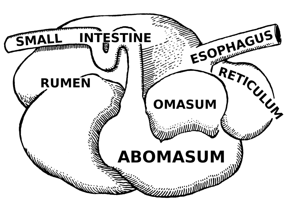
A typical ruminant stomach consist 4 chambers-
1. Rumen 2. Reticulum 3. Omasum 4. Abomasum.
The first three chambers (Rumen, Reticulum and Omasum) arise from the oesophagus and only the fourth, abomasum is an actual derivative of the stomach.
1. Rumen: Ruminants swallow partly chewed food into the first enlarged chamber – Rumen. Here the food is moistened and churned, mixing with symbiotic bacteria. Cellulase is produced from anaerobic bacteria that cleave the cellulose molecules into simple carbohydrates.
2. Reticulum (Honeycomb chamber): The wall lining is honey combed by ridges and deep pits. The inner wall is lined by mucous membrane. Small masses (cuds) are formed and chewed again and swallowed into the rumen.
3. Omasum (Psalterium): Thoroughly masticated and finely ground cud is followed into the rumen and passes into the 3rd chambers-the omasum. The mucous membrane is raised up into numerous longitudinal leaf-like folds.
4. Abomasum (Rennet): The abomasums have smooth vascular and glandular mucous membranes. The food material is processed by the usual digestive enzymes. This portion is the true glandular stomach with gastric gland.
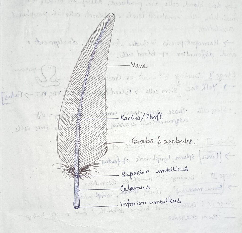
3.c) Describe the structure of a typical feather with a suitable diagram.
Ans→ A typical feather consists of – 1.) Axis/Main stem 2.) Vane/Vexillum
1.) Axis: Axis is divided into:– (I)Calamus & (II)Rachis
(I) Calamus: Proximal lower portion.
- Hollow, tubular and semi transparent.
- Base of the calamus is inserted into a pit or epidermal follicle of the skin.
- At the lower end of the quill has a small opening called inferior umbilicus.
- Superior umbilicus present at junction of the quill and rachis on the ventral surface.
(II) Rachis/Shaft: Distal upper portion of the axis.
- The rachis or the shaft forms the longitudinal axis of the vane.
- Longitudinal furrow (umbilical groove bear along the inner/ventral surface of rachis of its length)
2.) Vane The expanded membranous part of the feathers is called the vane /the vexillum.
- It is divided into two unequal lateral halves.
- Vane is formed by a series of the numerous parallel and closely spaced, delicate, thread structures, the barbs.
- Barbs are present at both side
- Each barb gives rise to a double row of many extremely delicate, oblique filaments called the barbules.
- The lower edge of the distal barbules is produced into minute hooklets, hamuli or barbicles.
Comparative Anatomy 2022
3.a) Briefly describe the structure of a contour feather of a bird with a labelled diagram.
Ans→Contour feathers are the large feathers that determine the shape and colour of the body.
Contour feather consists of – 1.) Axis/Main stem 2.) Vane/Vexillum
1.) Axis: Axis is divided into:– (I) Calamus & (II)Rachis
(I) Calamus: Proximal lower portion of the axis.
- Hollow, tubular and semi transparent.
- Base of the calamus is inserted into a pit or epidermal follicle of the skin.
- At the lower end of the quill has a small opening called inferior umbilicus.
- Superior umbilicus present at junction of the quill and rachis on the ventral surface.
(II) Rachis/Shaft: Distal upper portion of the axis.
- The rachis or the shaft forms the longitudinal axis of the vane.
- Longitudinal furrow (umbilical groove bear along the inner/ventral surface of rachis of its length)
2.) Vane The expanded membranous part of the feathers is called the vane /the vexillum.
- It is divided into two unequal lateral halves.
- Vane is formed by a series of the numerous parallel and closely spaced, delicate, thread structures, the barbs.
- Barbs are present at both side
- Each barb gives rise to a double row of many extremely delicate, oblique filaments called the barbules.
- The lower edge of the distal barbules is produced into minute hooklets, hamuli or barbicles.
3.b) Explain the tripartite concept of kidney organisation in vertebrates with a diagram.
Ans→ The tripartite concept of kidney organization in vertebrates is a model describing the development and organization of kidneys across different stages and types of vertebrates. This concept divides the kidney into three regions based on embryological development : the pronephros, mesonephros, and metanephros.
Pronephros:
- Location: Located anteriorly (towards the head) in the embryonic body.
- Development: It is the first kidney to develop in vertebrate embryos.
- Structure: Consists of a few pairs of nephrons.
- Function: In most vertebrates, the pronephros is non-functional or only temporarily functional. In some primitive fish (e.g., lampreys), it functions as the main excretory organ during the larval stage.
- Regression: In higher vertebrates, it degenerates and is replaced by the mesonephros.
Mesonephros:
- Location: Located in the mid-body region.
- Development: Develops after the pronephros, from the intermediate mesoderm.
- Structure: Comprises multiple nephrons that are more complex than pronephros.
- Function: Serves as the main excretory organ in fish and amphibians throughout life and in the embryos of higher vertebrates (reptiles, birds, and mammals).
- Regression: Functions during early development and is later replaced by the metanephros. However, parts of it contribute to reproductive structures (e.g., the Wolffian duct).
Metanephros:
- Location: Located posteriorly (towards the tail) in the body.
- Development: It is the last to develop, originating from the ureteric bud and the metanephric mesenchyme.
- Structure: Consists of numerous nephrons, forming a more complex and efficient excretory system.
- Function: Becomes the permanent kidney in reptiles, birds, and mammals.
- Efficiency: Metanephros is highly efficient in filtration and urine production in terrestrial life.
This tripartite concept illustrates the evolutionary and developmental progression of the vertebrate kidney, highlighting the increasing complexity and functionality from the pronephros to the metanephros.
3.c) Compare the structure of stomach of birds and mammals.
Ans→ Comparing the structure of the stomach in birds and mammals involves their anatomical and functional differences.
1.)Chambers:
- Birds: The avian stomach is divided into two main chambers: the proventriculus (glandular stomach) and the gizzard (muscular stomach).
- Mammals: Most mammals have a single-chambered stomach, though some, like ruminants (cows, sheep), have a multi-chambered stomach (rumen, reticulum, omasum, abomasum).
2.) Function of Chambers:
- Birds: The proventriculus secretes digestive enzymes and acids to break down food. The gizzard uses muscular contractions to grind food.
- Mammals: The single-chambered stomach in non-ruminants combines both chemical digestion (via enzymes and acid) and mechanical digestion (via muscular contractions).
3.) Mechanical Digestion:
- Birds: The gizzard is highly specialized for grinding, containing ingested stones that help in breaking down the food.
- Mammals: Mechanical digestion occurs through peristaltic movements of the stomach walls.
4.) Chemical Digestion:
- Birds: The proventriculus is the primary site of chemical digestion with enzyme and acid secretion.
- Mammals: The entire stomach lining secretes gastric juices, with parietal cells producing HCl and chief cells secrete pepsinogen.
5.) Layers:
- Birds: The proventriculus has a thick glandular mucosa for enzyme secretion, while the gizzard has a thick muscular layer and a tough, keratinized lining.
- Mammals: The stomach wall consists of mucosa, submucosa, muscularis, and serosa, with mucosal glands.
6) Adaptations for Diet:
- Birds: The gizzard’s structure is particularly adapted to a diet that often includes hard seeds and other tough materials requiring mechanical breakdown.
- Mammals: Stomach structure varies with diet; carnivores have elastic stomachs for large meals, while herbivores have more complex stomachs.
7.) Transit Time:
- Birds: Food moves relatively quickly from the proventriculus to the gizzard, with the grinding process.
- Mammals: Food stays in the stomach for a longer time, allowing mixing and breakdown by stomach acids and enzymes before moving to the small intestine.
