Answers of long question from PYQ Paper Animal Physiology of Semester IV of the years 2019, 2021 & 2022 respectively.
Animal Physiology 2019
3.a) Schematically describe the phenomenon of chloride shift. “Cl- content of RBCs in venous blood is significantly greater than that of arterial blood” -explain?
Ans→ It is the movement of Cl– in exchange with HCO3– across the RBC membrane.
Chloride Shift Phenomenon:
- CO₂ from the tissue entering the blood and then diffuses into RBC.
- CO₂ in RBC rapidly hydrated in the in the presence of carbonic anhydrase enzyme to form H₂CO₃.
- H₂CO₃ dissociates into H+ and HCO3–.
- HCO3– concentration in RBC increases and some of the HCO3– diffuses out to the plasma.
- In order to maintain electrical neutrality, chloride ions (Cl–) migrate/exchange from the plasma into the RBC.
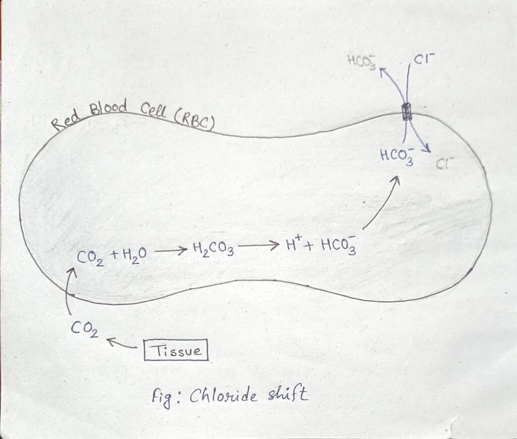
When the blood is deoxygenated (venous blood) in the circulation, CO₂ from tissues diffuses to RBC and forms HCO3– ion. This Heo ion diffuses out from RBC in exchange of Cl– ion to maintain electrical neutrality, ie, chloride shift takes place. So, Cl– content in RBC is more than arterial blood.
b) Briefly describe the hormonal regulation of digestion with suitable diagrams. What are the functions of bile?
Ans→ Gastrin: Produced by the stomach lining in response in presence of food. Gastrin stimulates the secretion of gastric acid and promotes gastric motility, preparing the stomach for digestion.
Secretin: Small intestine release secretin in exposure to acidic chyme come from the stomach. Secretin stimulates the pancreas to release bicarbonate ions, which neutralize the acidic chyme entering the small intestine.
Cholecystokinin (CCK): Secreted by cells in the small intestine in response to the presence of fats and proteins. CCK stimulates the gallbladder to release bile, which helps in the emulsification and digestion of fats. CCK also stimulates the pancreas to release digestive enzymes to further breakdown of fats, proteins, and carbohydrates.
Ghrelin: Known as the “hunger hormone” , produced primarily by the stomach when it’s empty. Ghrelin stimulates appetite and promotes food intake by acting on the hypothalamus in the brain, increasing feelings of hunger and initiating the process of digestion.
These hormones work together to ensure the efficient breakdown and absorption of nutrients.
c) What is cardiac cycle? How much time is required to complete one cardiac cycle? Describe in detail the mechanism of cardiac cycle.
Ans→ The cardiac cycle is the sequence of events that occurs in the heart during one heartbeat, involving the contraction (systole) and relaxation (diastole) of the atria and ventricles to pump blood throughout the body.
One cardiac cycle typically takes about 0.8 seconds in a healthy adult at rest.
Cardiac cycle completes in two phases –
Auricular Phase (0.8 sec):
Auricular Systole (0.1 sec): Atria contracts, pushing blood into the ventricles.
Auricular Diastole (0.7 sec): Atria relaxes and fills with blood from the veins.
Ventricular Phase (0.8 sec)
Ventricular Systole (0.3 sec):
- Isometric Contraction Period (0.05 sec): Ventricles start to contract with closed valves.
- Maximum Ejection Period (0.11 sec) : Ventricular pressure exceeds atrial pressure, forcing blood out.
- Reduced Ejection Period (0.14 sec) : Ventricular contraction slows, but blood continues to be ejected.
Ventricular Diastole (0.5 sec):
- Proto-Diastolic Period (0.04 sec): Ventricles relax, pressure drops, and semilunar valves close.
- Isometric Relaxation (0.08 sec): Ventricles continue to relax with all valves closed.
- Fast Rapid Filling Phase (0.113 sec): AV valves open, allowing blood to rush into the ventricles.
- Slow Filling Phase (0.167 sec) : Blood flow into ventricles decreases.
- Last Rapid Filling Phase (0.1 sec) : Atria contract again, completing ventricular filling.
Animal Physiology 2021
3.a) What is a cardiovascular centre? Where is it located? Describe how the cardiovascular centre regulates BP.
Ans→ The cardiovascular centre (or cardiovascular control centre) is a region in the brain that regulates heart rate and blood vessel diameter, thereby controlling blood pressure and blood flow.
Location: The cardiovascular centre is located in the medulla oblongata, which is part of the brainstem.
Cardiovascular centre regulates blood pressure (BP) by several steps –
Detection of BP Changes:
Baroreceptors in the carotid sinuses and aortic arch sense pressure changes. Chemoreceptors in the carotid and aortic bodies detect blood chemistry changes (O2, CO2, pH).
Signal Integration:
Signals from receptors are sent to the cardiovascular centre in the medulla oblongata.
Response Mechanisms:
1.) Sympathetic Activation (low BP response):
- Increases heart rate and contractility.
- Causes vasoconstriction, raising peripheral resistance.
2.) Parasympathetic Activation (high BP response):
- Decreases heart rate.
- Causes vasodilation.
Hormonal Regulation:
- Epinephrine and Norepinephrine: Increase heart rate, contractility, and vasoconstriction.
- Antidiuretic Hormone (ADH): Increases blood volume.
- Renin-Angiotensin-Aldosterone System (RAAS): Increases blood volume and causes vasoconstriction.
Feedback Mechanism:
Operates through negative feedback to maintain BP within an optimal range.
b) How do you differentiate Bohr effect and Haldane effect? Describe the process of transportation of metabolic CO₂ to the Lungs with a suitable diagram.
Ans→
| Bohr Effect | Haldane effect |
| 1. Describes how increased levels of carbon dioxide (CO2) and hydrogen ions (H+) in the blood reduce haemoglobin’s affinity for oxygen (O2). | 1. Describes how oxygenation of blood in the lungs reduces haemoglobin’s affinity for carbon dioxide and hydrogen ions. |
| 2. Facilitates oxygen release in tissues where CO2 concentration is high. | 2. Facilitates CO2 release in the lungs where oxygen concentration is high. |
| 3. Shifts the oxygen-haemoglobin dissociation curve to the right. | 3. Shifts the oxygen-haemoglobin dissociation curve to the left. |
Production of CO₂ in Tissues: Metabolic CO₂ is produced as a byproduct of cellular respiration.
Transport in Blood: CO₂ is transported in the blood to the lungs in three main forms:
- Dissolved in Plasma: About 5-10% of CO₂ is transported dissolved directly in the plasma.
- Bound to Haemoglobin: Approximately 20-30% of CO₂ binds to haemoglobin to form carbaminohemoglobin.
- As Bicarbonate Ions (HCO₃⁻): The majority (60-70%) of CO₂ is transported as bicarbonate ions. CO₂ diffuses into red blood cells, where the enzyme carbonic anhydrase catalyzes its conversion to carbonic acid (H₂CO₃). Carbonic acid quickly dissociates into bicarbonate ions (HCO₃⁻) and hydrogen ions (H⁺). Bicarbonate ions then diffuse out of red blood cells into the plasma, and chloride ions (Cl⁻) move into the red blood cells to maintain electrical neutrality, a process known as the chloride shift.
By these processes metabolic CO₂ transported into the Lungs.
c) Write about the process of fibrinolysis.
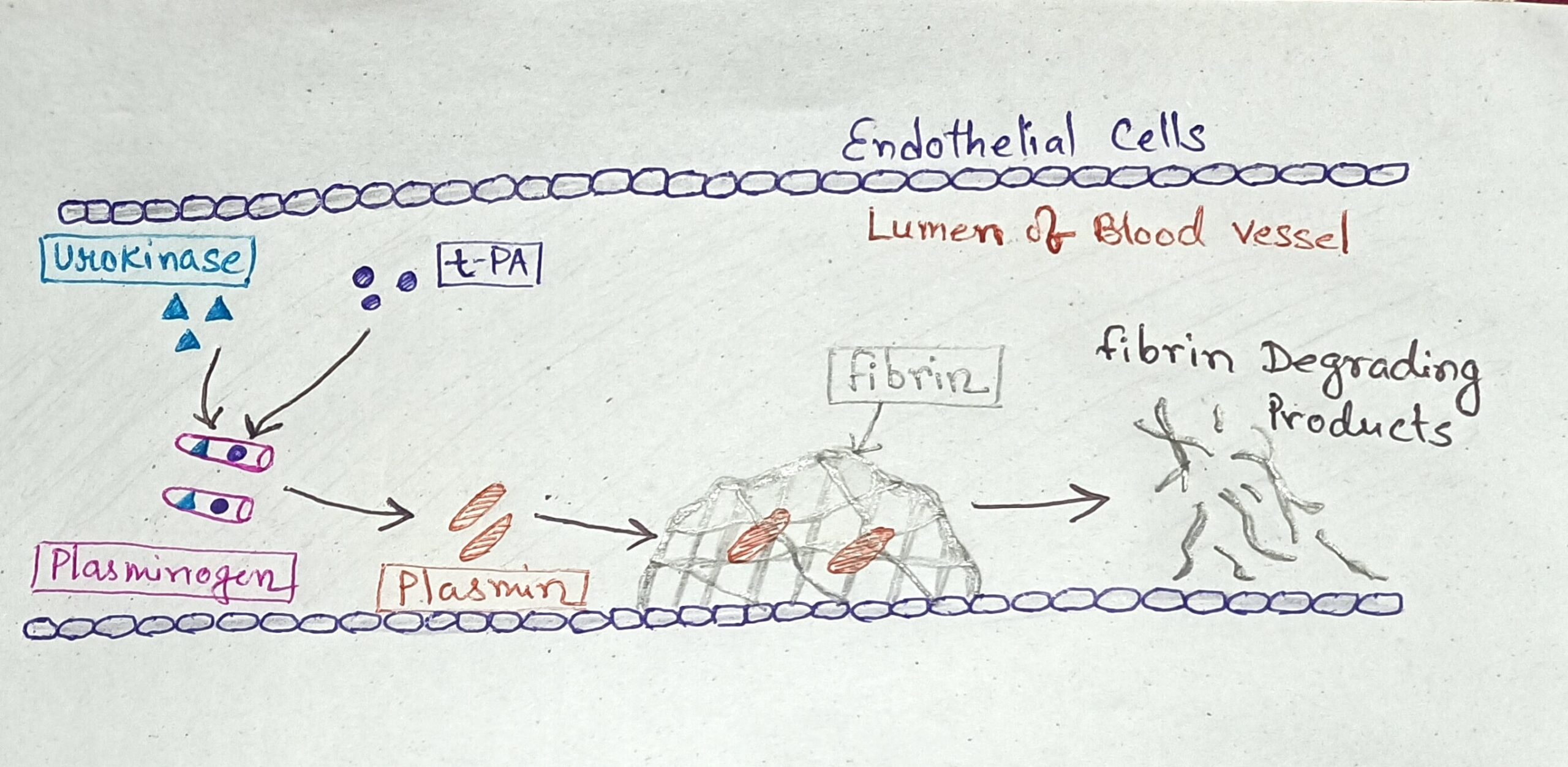
Ans→ Fibrinolysis is the physiological process that breaks down blood clots inside the blood vessel. And the process is controlled by the actions of various cofactors, inhibitors and receptors.
1.) Plasminogen Activation: The activation of plasminogen to plasmin is the central event in fibrinolysis. The activation is catalyzed by tissue plasminogen activator (tPA) and urokinase-type plasminogen activator (uPA).
2.) Formation of Plasmin: Once activated, plasminogen converts into plasmin, an active enzyme that can degrade fibrin.
3.) Degradation of Fibrin: Plasmin enzymatically cleaves the fibrin mesh into soluble fragments called fibrin degradation products (FDPs), effectively dissolving the clot.
4.) Regulation of Fibrinolysis: Plasminogen Activator Inhibitors (PAIs): These inhibit tPA and uPA, thereby regulating plasminogen activation. PAI-1 is the primary inhibitor and Alpha-2 Antiplasmin.
Resolution of Clot: As fibrin is degraded, the clot structure weakens and dissolves, restoring normal blood flow and vessel integrity.
Animal Physiology 2022
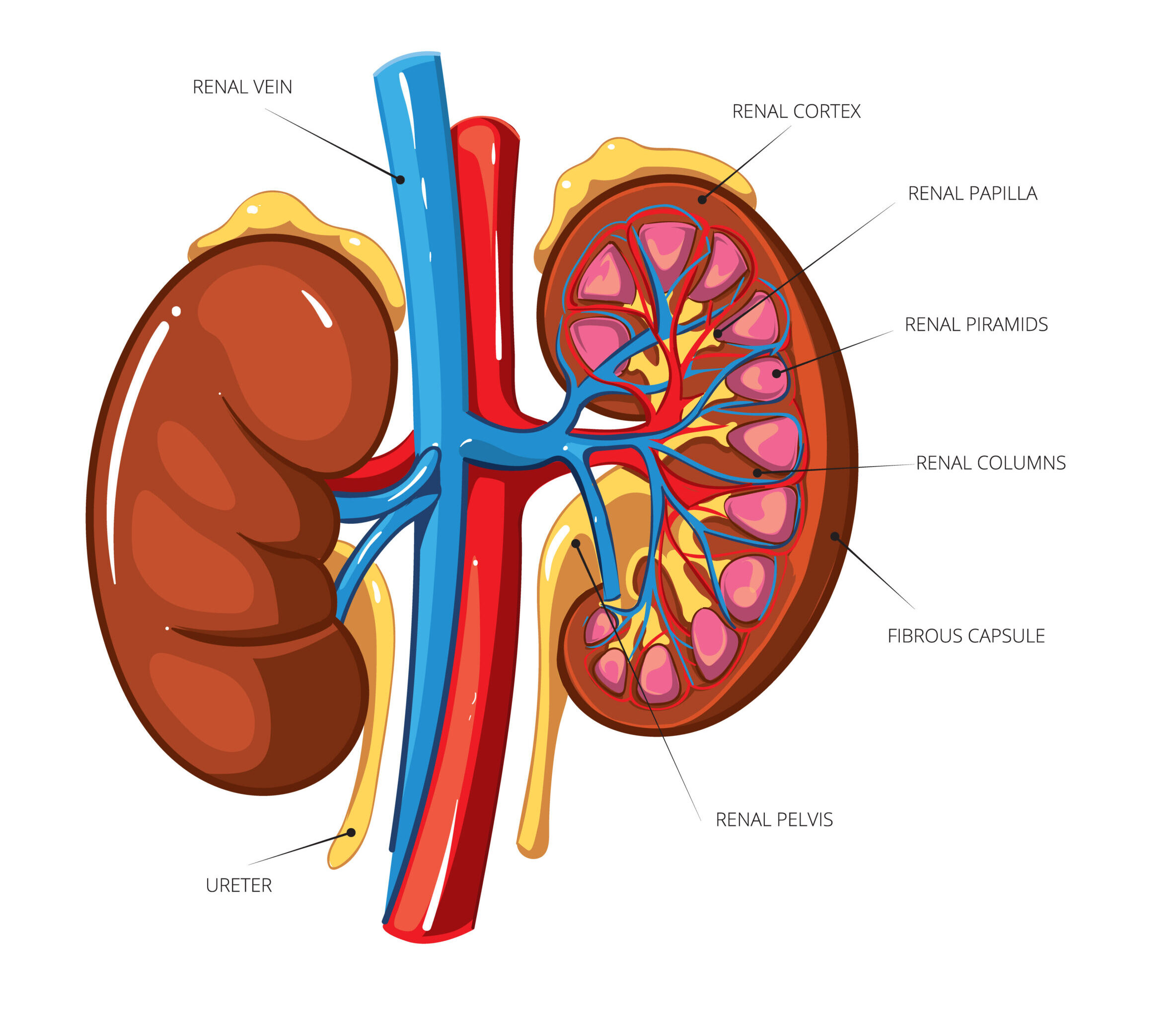
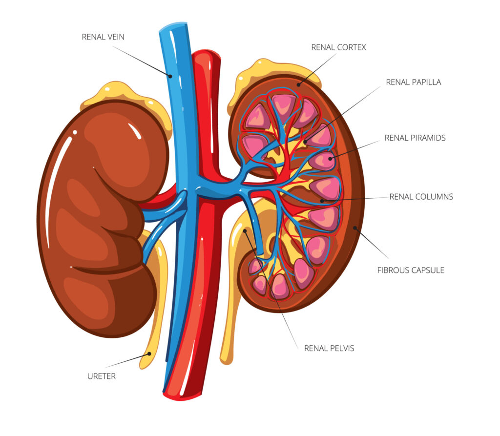
3. a) Draw and label the L.S of kidney. Differentiate Cortical and Juxtamedullary nephron.
Ans→
| Cortical Nephron | Juxtamedullary Nephron |
| 1. Glomerulus is in the upper region of the cortex. | 1. Glomerulus is near the junction of cortex and medulla. |
| 2. Includes 85% of nephrons. | 2. Includes 15% of nephrons. |
| 3. Glomerulus size is small. | 3. Glomeruli size is large. |
| 4. Rate of filtration is slow. | 4. Rate of filtration is high. |
| 5. Loop of Henle is small upto the outer layer of medulla. | 5. Loop of Henle is long and deep into the medulla. |
| 6. Contains a reduced network of vasa recta. | 6. Contains a large network Vasa recta. |
| 7. Major function – excretion of waste products in urine. | 7. Major function – concentrate the urine. |
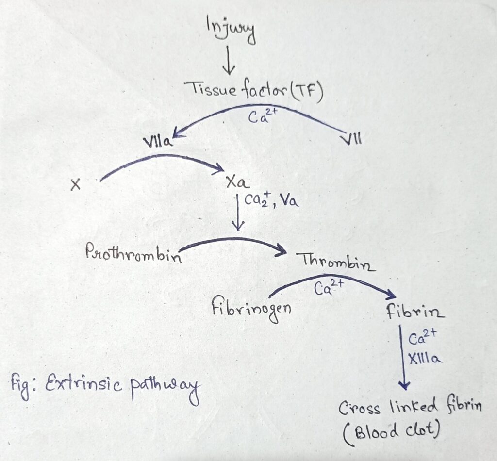
b) Schematically describe the extrinsic pathway for initiating blood clotting with special reference to the role Ca++.
Ans→ The extrinsic pathway of blood clotting is a shorter and occurs rapidly if trauma or injury is severe.
- When the vascular system is injured, Tissue factor (TF), a membrane-bound glycoprotein, is exposed at the site of injury.
- Tissue factor binds to circulating Factor VII, converting it to its active form, Factor VIIa. The TF-FVIIa complex, in the presence of Ca2+, activates Factor X by cleaving it to form its active enzyme, Factor Xa.
- Once Factor Xa is activated, it combines with factor V in the presence of Ca2+ to activate Prothrombin into Thrombin with the help of Prothrombinase and Ca2+.
- Ca2+ are necessary for the formation and function of the prothrombinase complex.
- Thrombin activates fibrinogen and then fibrin with Ca2+ ion and factor XIIIa forms cross linked fibrin or network of fibrin resulting blood clot.
[[ In summary, calcium ions (Ca++) play a critical role throughout the extrinsic pathway by:
- Facilitating the binding of FVII to tissue factor and its activation to FVIIa.
- Supporting the activation of FX and FIX by the TF-FVIIa complex.
- Enabling the formation and function of the prothrombinase complex.
- Ensuring the proper conversion of prothrombin to thrombin, leading to fibrin clot formation. ]]
c) What is thermogenesis? Describe the countercurrent system of temperature control in the human body.
Ans→ Thermogenesis is the process of dissipation of energy through the production of heat in an organism. [ Occurs in brown adipose tissue, skeletal muscle ]
When blood is flowing from the body core to the periphery (like legs & feet) it carries heat that can be readily lost through the skin. However, the vein returning blood to the body core lies alongside the artery. As the flow of warmer arterial blood and cooler venous blood encounters each other, exchange of heat takes place. Heat moves by conduction from the warmer arterial blood to the cooler venous blood. Even though the arterial blood cools on its way to the feet, the venous blood recovers heat from the arterial blood. This countercurrent heat exchange from warmer arterial blood to the cooler venous blood helps from losing heat through the peripheral skin.
